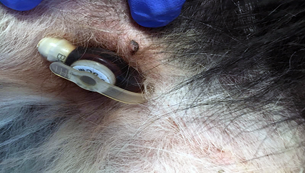
Percutaneously placed low-profile cystostomy tube
New Urinary Diversion Procedure For Dogs
We are very happy to offer percutaneously-placed, low-profile cystostomy tubes for management of obstructive urinary disease in dogs. Urinary diversion is a term used to described a procedure which diverts urine from the normal urinary apparatus to the external environment. Obstructive urinary disease is common in dogs and can occur due to benign or malignant, structural or functional uropathies. Depending on the underlying disease process, urinary diversion may be required for short-term (days-weeks) or long-term (months-years-indefinitely) management.
The most common location of urinary obstruction in dogs is the lower urinary tract with several disease processes potentially leading to obstruction at the level of the bladder / vesicourethral sphincter or urethra. Common structural pathologies leading to urinary obstruction which may require urinary diversion include transition cell carcinoma, prostatic carcinoma and granulomatous urethritis. Functional diseases which may lead to the requirement of urinary diversion include neurogenic bladder dysfunction (commonly associated with intervertebral disc disease and other structural vertebral or degenerative lesions) and detrusor-urethral (reflex) dyssynergia.
The most commonly performed procedures for urinary diversion for management of obstructive uropathies include urethral stenting and surgical placement of permanent cystostomy tubes. Average survival time after urethral stenting in dogs with transitional cell carcinoma is 2-3 months. Urinary incontinence occurs in approximately 50% of dogs following stent placement and clinical signs often recur due to stent obstruction by invasion / growth of the tumour. The placement of permanent cystostomy tubes has typically involved surgical intervention with the bladder approached and the tube placed via coeliotomy. While surgical placement of cystostomy tubes is effective, it is invasive and the large balloon / foley catheters typically used are cumbersome for owners to manage at home and are aesthetically unappealing.
SCVS are very happy to offer minimally-invasive, percutaneously-placed, low-profile cystostomy tubes for short, medium and long-term management of obstructive uropathies in dogs. We are not aware of any other centre in the UK offering this novel technique. The technique used avoids the need for surgical intervention and opening of the abdomen, which may be appealing to owners particularly in patients already diagnosed with a neoplastic disease (e.g. TCC) and where pursing a surgical intervention may not be perceived as an attractive option for management. When used for long-term management of urinary obstruction due to neoplastic disease, placement of a low-profile cystostomy tube offers the advantage of avoiding post-procedural incontinence. As the relatively short survival time post-stenting is often due to stent invasion with the growing tumour, this is obviously not a concern with cystostomy tube placement and as such, may lead to longer survival times. The benefit of percutaneously placement is that invasive surgery can be avoided and the use of low-profile tubes are much more visually appealing when compared to the larger ‘Foley-type’ catheters; the low-profile tubes are often not visible when dogs are in a standing position. Owners also report the tubes to be very well tolerated and easy to manage at home as no bandaging is required.
Briefly, the procedure for placement is as follows; the patient is anaesthetised and placed in dorsal recumbence. The ventral abdomen is clipped and aseptically prepared. An over-the-needle catheter is placed percutaneously into the bladder. A guide-wire is inserted into the bladder, through the catheter and the catheter is then removed. A graduated, peel-away dilator / introducer is inserted through the body wall and into the bladder lumen over the guide-wire. The guide-wire is removed and the low-profile cystostomy tube is insert via the peel-away introducer and held in place by inflation of a small balloon within the bladder lumen. The patient is recovered and hospitalised for at least 24 hours to allow initial seal formation around the stoma site.
The technique can be used for short, medium and long-term management of many diseases leading to partial or complete urinary obstruction. Examples include transitional cell carcinoma and prostatic carcinoma, neurogenic bladder dysfunction secondary to structural neurological disease, management of overflow incontinence due to degenerative neurological disease and detrusor-urethral dyssynergia. Please do not hesitate to contact us to discuss any potential cases which you feel may be appropriate or benefit from this procedure. We are always happy to discuss cases in advance of referral to determine suitability and further discuss options.
More from Southern Counties Veterinary Specialists

 7 years ago
  2660 views

 2 days ago