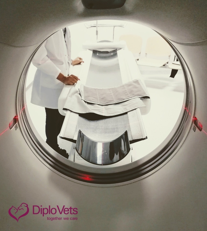
View from inside CT scanner looking out
Cone Beam Or Conventional CT - Which Is Best For My Practice?
All CT systems have one thing in common: they improve the quality of imaging diagnostics and expand therapy options. With an increasing demand for CT systems in the veterinary market, many veterinarians are faced with the question of which computed tomography system is best for their practice.
Not all CT scanners are the same
Veterinarians are essentially faced with the decision between two types of scanners: the multislice CT (also called Conventional CT) or the Cone Beam CT (CBCT, Digital Volume Tomography). Whether a Cone Beam or a Conventional CT system is better suited to your requirements depends on the individual range of diagnostics and treatment and the specialisation of your team. In both cases, modern CT units are characterised by high image quality, but they differ fundamentally in terms of their possible applications.
Veterinary imaging experts agree that Conventional CT is the recommended choice for generalised medicine and surgery practice with its wide range of applications.
However a closer look at the advantages of Cone Beam CT would highlight its specialised advantages.
Cone Beam CT - leading in smallest structures and dentistry
A Cone Beam CT provides significant advantages in small anatomical structures with high tissue density like head structures or joints. In these areas, a Cone Beam CT creates superimposition-free, detailed 3-D reconstructions and clearly displays even fine structures of the examined region - in dentistry a considerable advantage over dental x-rays.
This leads to the following advantages in imaging and possible applications for a Cone Beam CT:
- Head structures: Increased spatial resolution leads to most detailed diagnostics in dentistry, oral and maxillofacial medicine
- Small pets and exotic animals: Cone Beam CTs are also suitable for head- and even whole body examinations of exotics or small pets like rabbits, guinea pigs or even of the size of a small rodent
- Joints: Cone Beam CT systems can also be used in orthopaedics, e.g. to display smallest joint structures
Apart from the imaging, a Cone Beam CT offers the following advantages:
- Lower radiation dose required than a Conventional CT system (a factor which may be insignificant in veterinary scanning)
- Lower purchase and energy costs
- Reconstruction software can be installed on common computers - therefore they do not require a workstation coupled to tomography. This reduces costs and space consumption and is saving reconstruction measures
The difference in the beam path and its effect on contrast
Contrast quality depends heavily on the straight beam path from the generator through the anatomy to the detector. If the radiation is scattered, the contrast resolution is reduced. High contrast is especially important in the thoracic and abdominal regions, where these subtle differences are usually the reason why CT is chosen as an imaging technique of choice.
Conventional CT systems have a collimated beam so that it runs as straight as possible and little scattered radiation is produced or detected. This is precisely the major difference of the cone beam technology: The x-rays from the Cone Beam CT are not collimated. The x-rays are forming a divergent, uncollimated cone, so that more scattered radiation is produced. This leads to a reduced soft tissue contrast resolution, especially in the thorax and abdominal region. As with X-rays, the larger the body part images, the greater the scatter. For example, using a Cone Beam CT for a large dog abdomen or thorax, the contrast would be lower than for a small pet or limb.
A Cone Beam CT is therefore not suitable for routine use in thoracic and abdominal regions or for soft tissue examinations.
Motion artefacts
In Cone Beam CT, the images of the field of view are acquired in one complete arc, whereas a Conventional CT produces numerous image slices, each of which requires a separate scan.
For this reason, a Cone Beam CT is more susceptible to motion artefacts than a slice CT: Even one motion artefact can reduce the overall image quality and can lead to the necessity of repeating the entire scan. Conventional CT systems are less affected by motion artefacts because in slice CT, only the slices with movement show artefacts while the other slice exposures are not affected.
Clear limitations of Cone Beam CT systems
When evaluating CT scanners, you should consider image quality as the most important factor.
If you are only working on facial, jaw and dental diseases or small pets, a Cone Beam CT saves costs and can still achieve the best quality in these regions.
But be aware: On the other hand, there are strong limitations of a Cone Beam CT, which should be clearly considered when it comes to soft tissue and body cavity examinations!
If your practice performs more than head structure scans or mostly thorax and abdominal CTs on small or large animals, you need to purchase a multislice CT, which offers the required high contrast and less susceptibility for motion artefacts, e.g. due to breathing.
You are therefore much more broadly positioned with a Conventional CT.
Many radiologists do not work regularly with Cone Beam CTs but only with multislice CTs.
If you have your images evaluated externally, e.g. by a teleradiology provider, Cone Beam images are usually only evaluated if Cone Beam CT scans are used in the appropriate area.
Do you need support?
DiploVets offers more than just diagnostics. We also understand us as a point of contact for questions and uncertainties that arise in connection with imaging.
Together we achieve the best diagnosis and treatment options.
Contact:
E [email protected]
W www.DiploVets.com – together we care
More from DiploVets
- What legal requirements do I have to consider before purchasing a CT system?
- Do I purchase a new or refurbished computed tomography?
- What considerations lead me to the appropriate provider for a CT system?
- Where can I find well-founded information on CT scanners?
- Are you prepared to implement a CT modality in your practice?

 3 years ago
  1082 views
