
Hallmarq Veterinary Imaging Adds Standing Equine Leg CT (slCT) To Advanced Imaging Portfolio
 5 years ago
  1466 views
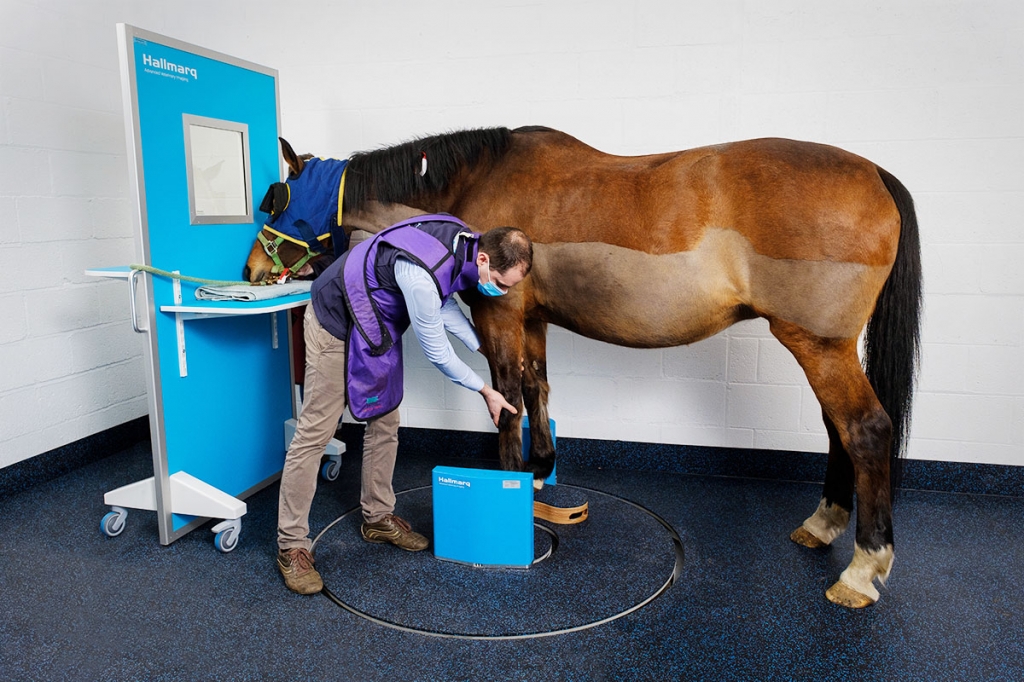
Guildford UK —To help vets quickly and safely diagnose the cause of equine lameness, Hallmarq Veterinary Imaging recently announced the addition of Standing Equine Leg CT (slCT) to its product range, increasing access to advanced imaging for all equine vet practices. Hallmarq is introducing the new slCT after achieving success imaging horses in collaboration with their UK-based clinical trial sites. Hallmarq Veterinary Imaging pioneered the development of standing MRI for horses and the new standing CT scanner uses a simple and safe design, with a unique low, flat platform for quick and easy entry and exit of the standing sedated horse.
The system uses a novel dual-concentric ring design which enables the detector plate to remain very close to the region of interest, thereby improving image quality. slCT is a good fit for equine practices wanting to step up to 3D imaging in the evaluation of their lameness cases. Hallmarq is one of the few companies to incorporate motion correction technology to better ensure high quality, clear images in the standing patient.
To aid in increasing access to this technology for more practices, Hallmarq considers the total cost of ownership in its design decisions, providing an affordable solution to most clinics. Their unique financial models and low start-up costs negate the need for upfront capital expenditure and their flexible monthly payment options take seasonality into account.
Bell Equine in Kent, Sussex Equine in Ashington, and Berkshire- based Donnington Grove Equine have been instrumental in the early trials of the novel system. Dr Elisabetta Giorio from Donnington Grove is enthusiastic about their involvement stating:
“It was really quite exciting to work with the Hallmarq team to help develop this system.”
Hallmarq has capitalised on over 20 years of experience with their unique Standing Equine MRI (sMRI) to create this additional tool for equine vets to fully evaluate and diagnose bony disease in the equine distal limb, including 3D visualisation of fractures. CT combines hundreds of x-rays to create a 3D digital image, which can then be viewed as thin slices from any direction, eliminating overlapping structures and revealing minute detail in bone and cartilage. slCT complements their sMRI which highlights soft tissue and metabolic changes and together they provide an even more powerful diagnostic aid, according to Giorio:
“The combination of MRI and CT was a useful tool to have and helped with surgical planning and decision making,”
The value of using both modalities together is one that Dr Alison Fairburn, Specialist in Diagnostic Imaging at Bell Equine Vets also recognises:
“It really complemented what the MRI had told us about the content and signal intensity and, in this case, helped with surgical planning, providing useful information on the bone margin.”
Hallmarq’s dedicated Research and Development team has been working in collaborative partnerships with their phase one clinical trial sites for the past year, imaging live horses to ensure that the system meets the image quality, robustness and efficiency levels required for use in clinical practice. As with their other systems, Hallmarq considers the practices’ total cost of ownership in its design decisions, providing a solution that is accessible to most clinics. Cone beam technology (CBCT) is ideal for distal limb imaging where it can detect non-displaced fractures, subtle changes in bone density and small osteophytic lesions without the expense, radiation risks and 3-phase power requirements of fan beam systems.
Dr Luis Rubio-Martinez, surgeon at Sussex Equine Hospital, is keen to test this technology further, adding:
“We are interested to see if the slCT can improve our ability to diagnose cartilage defects in coffin joints and fetlocks.”
As installations continue, the new Hallmarq system is now up and running at Rainbow Equine Hospital, North Yorkshire.
Adding slCT to their state-of-the-art diagnostics suite was an obvious next step forward for the practice, as Dr. Jonathon Dixon, member of the referral team and European Specialist in Veterinary Diagnostic Imaging, explains:
“We are really excited to put into practice the development so far to help facilitate the move into second phase clinical trials and offer our customers a world first.”
Further information on Hallmarq's Standing Equine Leg CT (slCT) is available at https://hallmarq.net/products-services/standing-equine-leg-ct/.
More from Hallmarq Veterinary Imaging
- Hallmarq Veterinary Imaging expands in-house production to meet customer demand
- Hallmarq installs its first zero-helium small animal 1.5T MRI machine in the US
- Hallmarq’s Vision CT campaign to receive VETTY Award® for excellence in animal health marketing at VMX 24
- Hallmarq Veterinary Imaging’s new US facility meets rising demand for equine MRI and CT systems
- Hallmarq Veterinary Imaging’s new US facility meets rising demand for equine MRI and CT systems
